
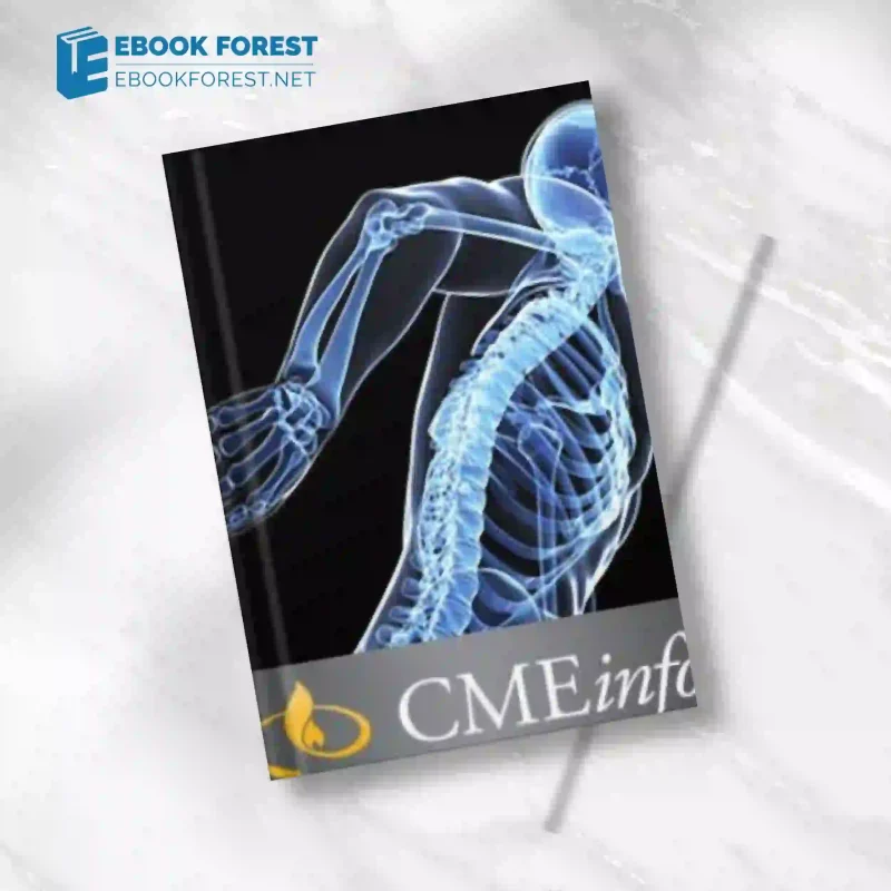
The UCSF Musculoskeletal MR Imaging activity from the Department of Radiology and Biomedical Imaging at the UCSF School of Medicine provides a thorough state-of-the-art update on many facets of musculoskeletal MRI. Assessment of cartilage lesions, emphasizing the most recent sequences that are available, is addressed. For these reasons, a differential diagnosis is provided along with a thorough evaluation of soft-tissue and bone cancers. A number of presentations cover relevant anatomy and pathology for each joint in addition to optimized methods for 1.5T and 3T MR imaging equipment. Useful information is focused on nerve entrapment, muscle and tendon diseases, and bone marrow.
The UCSF Musculoskeletal MR Imaging activity provides an extensive, up-to-date overview of the various facets of musculoskeletal magnetic resonance imaging. Assessment of cartilage lesions, emphasizing the most recent sequences that are available, is addressed. For these reasons, a differential diagnosis is provided along with a thorough evaluation of soft-tissue and bone cancers. A number of presentations cover relevant anatomy and pathology for each joint in addition to optimized methods for 1.5T and 3T MR imaging equipment. Useful information is focused on nerve entrapment, muscle and tendon diseases, and bone marrow.
Diagnostic radiologists, orthopedic surgeons, sports medicine doctors, and MRI technologists who wish to develop their understanding of musculoskeletal MRI from a basic or intermediate level are the target audience for this activity.
Subjects & Presenters
MRI of the post-operative shoulder, biceps, tendon, and elbow
Dr. Lynne S. Steinbach
Multimodality Imaging of Anterior Cruciate Ligament Graft Reconstruction Anterior Knee Pain Imaging
Dr. Daria Motamedi
Ankle MRI
Doctor Thomas M. Link, PhD
What Knee MRI Information Does the Surgeon Want to Know?
Shoulder MRI: What Is and Is Not Important?
Benjamin C. Ma, M.D.
Cartilage in Relation to Osteochondritis
Hip MRI: Case-Based Bone Tumor Imaging
both radiofrequency ablation and bone biopsies
Doctor Thomas M. Link, PhD
Impingement and Rotator Cuff
Overhead throwing athletes’ knee meniscus injuries and patterns of bone marrow edema
Christine Chung, M.D.
MSK Injections: Image-Guided Diagnostic and Therapeutic Procedures
MRI Following Joint Replacement
John Feller, M.D.
Faculty Questions and Discussion
Chair of Faculty Courses
Thomas M. Link, MD, PhD, Chief of Musculoskeletal Imaging and Professor of Radiology
VISITOR DEFICIENCY
Christine Chung, MD, UC San Diego School of Medicine Professor of Radiology
John Feller, MD American Medical Center in Shanghai, China Desert Medical Imaging Indian Wells, California
UCSF Department of Education
Chief of Sports Medicine and Shoulder Service and Associate Professor of Orthopaedic Surgery, C. Benjamin Ma, MD
Radiology Assistant Professor Daria Motamedi, MD
Professor of Radiology and Orthopaedic Surgery Lynne S. Steinbach, MD
Learning Goals
Following completion of this task, participants will be able to: Connect the dots between the orthopedic surgeon’s perspective and the results of MR imaging of the shoulder and knee.
Provide musculoskeletal imaging procedures that are optimized, particularly those that suppress metal hardware in patients undergoing joint replacements.
Determine the clinical relevance of any abnormalities found on an MRI of the joints, muscles, bone marrow, and cartilage.
Identify typical bone cancers using a relevant differential diagnosis
Recognize the anatomy of the elbow, shoulder, hip, knee, and ankle from MRI scans.
Suggest joint injections for diagnostic and therapeutic purposes based on imaging guidance.
Recognize the uses, precautions, and methods for radiofrequency ablation and bone biopsies.
Gratitude
The Accreditation Council for Continuing Medical Education (ACCME) has granted UCSF School of Medicine its accreditation to offer continuing medical education to doctors.
Release Date of the Series: May 16, 2014
File size total: 4 GB
See also: UCSF Breast Imaging and Digital Mammography 2014 Videos


Reviews
There are no reviews yet.